Bioelectricity: Electric excitability and action potential
 What is an action potential?
So far we have assumed a passive cell model, i.e. a model in which the membrane properties, and consequently the membrane potential, are immutable. However, the membrane potential in electrically excitable cells such as nerve cells and muscle cells can temporarily change in response to a stimulus at a time scale of a few milliseconds. Such an action potential (for nerve cells in English also called spike) mainly appears to be triggered by a temporary change in the conductivity of the membrane for potassium and sodium ions. It is a local response to a stimulus that, however, is transmitted to the surrounding part of the membrane and thus leads to a propagated signal.
The stimulus may be a temporary decrease of the membrane potential relative to the resting membrane potential, a phenomenon which is called hyperpolarization, but this will only lead to a gradual change of the membrane potential and the response to the stimulus is proportional to the magnitude of the stimulus.
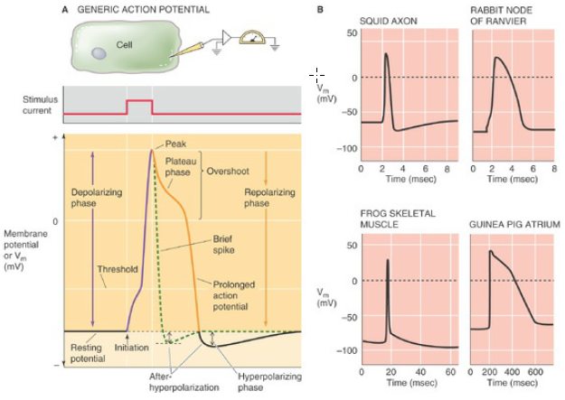
Much more interesting is a temporary increase in the membrane potential with respect to the resting membrane potential, a phenomenon that is called depolarization. In case of a not too large stimulus, the membrane potential gradually adapts to the new conditions, and the response is proportional to the magnitude of the stimulus. But spectacular is the effect when the stimulus makes the membrane potential pass a certain limiting value, the so-called potential threshold or firing threshold. Then an action potential is created: the membrane potential increases in a very short time from a negative to even a positive value (the part of the action potential above 0 mV is called the overshoot) and then in the repolarization phase slowly returns back to its original resting membrane potential. During this return it may even happen that the membrane potential temporarily becomes lower then the resting membrane potential (undershoot or afterhyperpolarization), but this depends on the type of the cell. In the figure below is shown the action potential as a result of a current injection for different tissues.

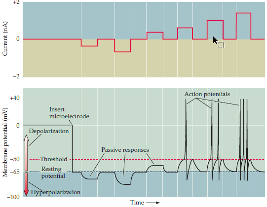
What is further worth noting, is the fact that the shape and the height of the action potential hardly depends on the strength of the stimulus: it is just a matter of stimulation that brings the membrane potential above the firing threshold or keeps it below this threshold which determines whether or not an action potential is triggered or not. It is an all-or-none phenomenon. But we will see that the strength of the stimulus affects the frequency of repetition of the action potential: a large stimulus translates into a closely spaced series of action potentials (spike train), while a mild stimulus leads to more space between action potentials. The figure below illustrates this.

In this section we will discuss the model that Hodgkin and Huxley developed for the generation of an action potential or a spike train in the fifties of the last century and for which they received the Nobel prize. They have created their model on the basis of measurements made on a giant nerve fibre (giant axon) of a squid. It is the standard example for modelling of action potentials in a cell: many of the other models are variations on the theme.


